How EEG Spike Detection Is Reshaping Diagnostics
Posted by Pabitra Giri
Filed in Health 210 views
Neurology Has a Workflow Problem — and AI Is Finally Solving It
There's a paradox sitting at the center of modern epilepsy diagnostics. The tools available to neurologists today are better than they've ever been. EEG hardware is more sensitive. Long-term monitoring capabilities have expanded. The clinical evidence base for seizure management has deepened. And yet one of the most fundamental bottlenecks in epilepsy care — the time and cognitive load required to accurately review EEG data — has remained largely unchanged for decades.
The math is straightforward and sobering. A patient admitted to an epilepsy monitoring unit might generate 24 to 72 hours of continuous EEG recording. That data contains the clinical answers the physician needs: where spikes are occurring, how frequently, what seizure patterns look like, whether events are focal or generalized. But extracting those answers requires expert review of a volume of data that exceeds what any single reviewer can process with consistent accuracy under typical clinical time constraints.
That gap — between the information available in the data and the practical capacity to extract it — is where diagnostic delays, missed events, and review inconsistencies enter the picture. It's a structural problem, and individual effort and expertise alone can't fully resolve it.
AI-assisted EEG analysis, specifically AI-powered eeg spike detection, is the most significant structural answer to that problem that clinical neurology has seen. Not because it replaces the neurologist, but because it fundamentally reshapes what the neurologist's time is spent on.
Understanding the Diagnostic Weight of EEG Spikes
To appreciate why AI-assisted spike detection matters clinically, it helps to understand what neurologists are actually looking for when they review EEG data for epileptic activity.
Epileptiform discharges — spikes, sharp waves, and spike-wave complexes — are the primary interictal markers that indicate epileptic potential in neural tissue. They appear between seizures, often during periods when patients are symptom-free, and they provide a window into the underlying epileptogenic activity that drives seizure generation.
The clinical value of identifying these discharges accurately is substantial and multidimensional.
Localization. The distribution and field of spike activity across the electrode array helps neurologists identify the brain regions involved in seizure generation. This localization is essential for surgical planning, treatment selection, and prognosis.
Syndrome identification. Different epilepsy syndromes produce distinct spike patterns. Childhood absence epilepsy, juvenile myoclonic epilepsy, focal cortical dysplasia, and temporal lobe epilepsy each have characteristic EEG signatures. Identifying these signatures correctly shapes the entire management pathway.
Treatment monitoring. Changes in spike frequency and distribution over time — in response to antiseizure medications or other interventions — provide objective markers of treatment response. Systematic tracking of spike activity requires systematic detection, which is precisely what AI makes more tractable.
Surgical candidacy. For patients who don't respond to medication, surgery may be the most effective path to seizure freedom. Accurate spike localization is a core input to the presurgical evaluation process. Missed or mislocalized spikes can misdirect the surgical workup in ways that matter enormously to patient outcomes.
The Technical Challenge Behind Accurate Spike Detection
Building an AI system that reliably detects spikes in real-world clinical EEG is a harder problem than it might appear. The variability in spike morphology is enormous — across patients, across scalp regions, across EEG recording conditions, and across the clinical populations that differ in age, medication status, and neurological background.
Artifacts compound the challenge. Electrode artifacts, movement artifacts, cardiac artifacts, and muscle activity can all produce waveform features that superficially resemble epileptiform activity. A system that can't reliably distinguish genuine spikes from artifact-generated mimics generates a false positive burden that undermines its clinical utility.
High-sensitivity detection is necessary but not sufficient. The system also has to be specific enough that the physician review workload is actually reduced rather than relocated. Getting this balance right requires training data that reflects the full complexity of real clinical EEG — including the messy, artifact-contaminated, variable recordings that characterize actual hospital data rather than curated research datasets.
How NeuroMatch Addresses the Detection Challenge
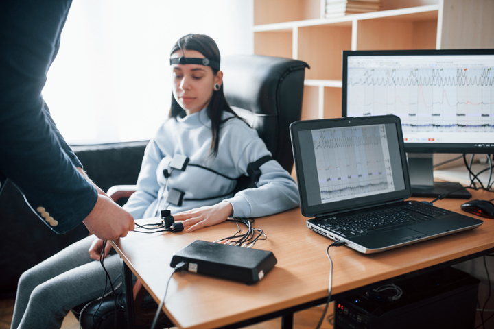
LVIS Corporation's approach to this problem is grounded in the depth and quality of the data used to develop and validate its detection algorithms. The eeg software powering NeuroMatch's spike detection capability was trained on thousands of hours of 19-channel EEG data — the standard clinical configuration used in hospital and monitoring settings across the United States.
Training at that scale, on data that reflects real clinical recording conditions, is what allows the system to handle the morphological variability and artifact complexity that simpler or less extensively trained systems struggle with. The result is detection performance that holds up in actual clinical deployment — not just in controlled validation studies.
The seizure detection capability works alongside spike detection, using deep-learning algorithms to identify seizure events and notify physicians within an hour. For ICU monitoring and epilepsy monitoring units where seizure identification speed directly affects intervention timing, that notification window is a meaningful clinical capability.
Critically, both features are FDA-cleared for clinical use in the United States. That clearance is the result of a rigorous regulatory process that validates the system's safety and effectiveness for its intended clinical use — and it's the standard that should be expected of any AI-assisted diagnostic tool deployed in US clinical settings.
Integration Into Clinical Workflow: The Design That Makes It Work
Technology that doesn't fit into clinical workflow doesn't get used — and when it doesn't get used, patients don't benefit. This is a consistent challenge in health technology adoption, and it's one that LVIS Corporation has addressed directly in how Neuromatch is designed.
The platform is built around physician-in-the-loop workflow rather than autonomous AI decision-making. When the system detects a spike event or seizure, it presents that detection to the reviewing physician as a candidate — something to be validated, confirmed, or adjusted based on clinical judgment. The AI does the pattern recognition work across the full dataset. The physician brings clinical expertise, contextual judgment, and final authority to the interpretation.
This design serves both clinical and regulatory requirements. It preserves physician autonomy and liability structures that clinical practice demands. It creates a documentation trail for AI-assisted detections and physician review decisions. And it ensures that the system's utility is genuinely additive — physicians aren't being asked to override a black box, they're being supported by an intelligent layer that makes their expertise more efficient.
Deployment Experience and Real-World Validation
NeuroMatch was launched in the United States in January 2025 and had already been successfully deployed in more than ten hospitals in South Korea prior to its US launch. That real-world deployment history is clinically meaningful — it means the platform's performance has been tested in actual hospital environments, across diverse patient populations and clinical settings, rather than only in controlled research conditions.
The ability to automate key aspects of EEG interpretation — spike identification, seizure flagging, workflow notification — streamlines clinical operations in ways that have practical consequences for how hospitals can scale their neurological diagnostic services. For institutions looking to expand EEG monitoring programs, manage growing patient volumes, or extend diagnostic capabilities to settings where specialist density is limited, this kind of automation is not a luxury — it's an enabling capability.
The Broader Implications for Neurological Diagnostics in the US
The trajectory of AI-assisted EEG analysis points toward a future in which the quality gap between well-resourced academic centers and community or rural settings narrows considerably. Accurate, validated, FDA-cleared eeg spike detection — available through a scalable software platform rather than capital-intensive hardware infrastructure — changes the accessibility equation for neurological diagnostics in meaningful ways.
For the roughly 3.4 million Americans living with epilepsy, many of whom receive care in settings that currently lack full specialist support, that change in accessibility has direct patient consequences.
See What NeuroMatch Can Do for Your Program
If you're evaluating AI-assisted EEG analysis tools for your neurology department, epilepsy monitoring unit, or hospital system, LVIS Corporation's NeuroMatch platform offers a combination of clinical depth, regulatory clearance, and workflow-integrated design that sets a high bar in the market. Visit lviscorp.com to learn more, review the platform's clinical evidence, and schedule a demonstration with the LVIS team.